
Introduction
-
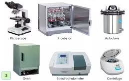
Histology laboratory equipment is essential for preparing and examining tissue samples for microscopic study.
-
These tools ensure proper fixation, processing, sectioning, and staining of tissues.
-
Microtomes and cryostats enable the cutting of thin, accurate tissue sections.
-
Tissue processors automate dehydration, clearing, and infiltration steps for uniform results.
-
Embedding centers and water baths support proper block orientation and section handling.
-
Staining equipment enhances tissue contrast for clear visualization under the microscope.
-
Microscopes are crucial for analyzing cellular details and identifying pathological features.
-
Fume hoods provide a safe workspace for handling hazardous chemicals used in histology.
-
Refrigerators, pH meters, and storage systems help maintain reagent quality and stability.
-
Proper maintenance of all equipment ensures consistent, accurate, and safe laboratory performance.
Laboratory Equipment
Microtome
Principle
A microtome advances a paraffin-embedded tissue block toward a sharp steel/ceramic blade at controlled thickness increments (1–20 µm), producing reproducible thin sections.
Uses
-
Cutting thin sections (typically 3–5 µm for routine H&E; 7–10 µm for special stains like Congo Red).
-
Essential for creating high-quality tissue ribbons without compression, chatter, or tearing.
-
Used for paraffin, resin, and cell blocks.
Maintenance Practices
1. Blade Care
-
Replace disposable blades once they show dullness (compression artifacts, chatter).
-
Handle blades with forceps; never with fingers.
-
Store unused blades in a dry environment to prevent rusting.
2. Cleaning
-
Clean microtome stage, clamps, and knife holder daily with xylene-free or non-corrosive disinfectant.
-
Remove wax residues with mild heat or xylene (according to lab SOP).
3. Lubrication & Mechanical Checks
-
Lubricate moving parts weekly (using manufacturer-recommended oils).
-
Check block holder alignment to avoid tilted sections.
4. Calibration
-
Verify thickness settings monthly with calibration blocks or plastic strips.
-
Confirm even advancement of handwheel.
Best Practices
-
Always lock the microtome when not in use.
-
Maintain room at 18–22°C and moderate humidity for optimal sectioning.
-
Avoid excessive force during block trimming.
Cryostat
Principle
A cryostat is a refrigerated chamber containing a microtome for cutting frozen tissue sections at −20°C to −30°C, preserving lipids, enzymes, and antigens.
Uses
-
Intraoperative rapid diagnosis (frozen sections).
-
Immunofluorescence (renal biopsies).
-
Lipid staining (Oil Red O), as lipids dissolve in paraffin.
-
Enzyme histochemistry requiring rapid freezing.
Maintenance Practices
1. Temperature Checks
-
Verify chamber temperature daily.
-
Maintain:
-
−20°C for soft tissues
-
−30°C for dense tissues like muscle
-
2. Blade Maintenance
-
Replace blades frequently due to rapid dulling from cold cutting.
-
Dispose of blades in puncture-proof sharps container.
3. Cleaning & Disinfection
-
Remove tissue debris after each use.
-
Perform weekly defrost cycles to remove frost buildup.
-
Use 70% ethanol or cryostat-safe disinfectants.
-
UV disinfection (if present) should be run at least once per day.
Best Practices
-
Keep chamber closed as much as possible to prevent ice accumulation.
-
Wear PPE—cryostat can aerosolize tissue particles.
-
Maintain separate cryostat for infectious specimens.
Tissue Processor (Automatic / Semi-automatic)
Principle
Automated movement of tissues through dehydration (alcohols), clearing (xylene/ substitutes), and paraffin infiltration.
Uses
-
Provides uniform processing for consistent tissue morphology.
-
Saves labor and reduces human error in multiple reagent transfers.
-
Ensures optimal infiltration for easy microtomy.
Maintenance Practices
1. Reagent Management
-
Monitor alcohol/xylene/paraffin levels daily.
-
Replace reagents based on cycles, color change, or impurity load.
-
Maintain logs for reagent changes for QA audits.
2. Chamber Cleaning
-
Clean retort with warm paraffin or alcohol-based cleaning solutions.
-
Check tubing for blockages or leaks weekly.
3. Calibration
-
Validate timing, pressure cycles, and temperature monthly.
-
Ensure paraffin bath remains at 2–4°C above melting point.
Best Practices
-
Use standardized cassette loading patterns to ensure even infiltration.
-
Avoid overloading retort—causes incomplete processing.
-
Monitor tissue size recommendations (usually <4–5 mm thickness).
Embedding Center (Embedding Station)
Principle
Maintains molten paraffin for embedding tissues into blocks, enabling correct orientation for sectioning.
Uses
-
Embedding tissues in paraffin with correct anatomical orientation.
-
Solidifying blocks on a cold plate for microtomy.
Maintenance Practices
1. Temperature Control
-
Paraffin reservoir: 58–60°C
-
Heated forceps wells: slightly above melting point
-
Cold plate: −5°C to −10°C
2. Cleaning
-
Remove paraffin spills immediately.
-
Weekly cleaning of dispensing nozzles to prevent clogging.
3. Paraffin Quality
-
Replace paraffin when discolored or contaminated.
Best Practices
-
Pre-warm cassette and mold to prevent bubbles.
-
Ensure tissue is fully surrounded by paraffin (no air pockets).
Staining Equipment (Manual Racks / Automatic Stainers)
Principle
Sequential application of dyes, buffers, differentiators, and bluing agents to produce contrast.
Uses
-
H&E staining (routine).
-
Special stains (PAS, Masson’s Trichrome, reticulin, silver).
-
Ensures reproducible staining for diagnostic quality.
Maintenance Practices
1. Daily Cleaning
-
Wash racks post-use with mild detergent.
-
Remove precipitates and dye residues.
2. Reagent Monitoring
-
Replace hematoxylin when oxidized.
-
Monitor alcohol and xylene purity.
-
Check expiration dates and clarity of solutions.
3. Calibration (Automatic Stainers)
-
Validate dip times, agitation patterns, and temperature settings.
-
Check robotic arms and slide holders.
Best Practices
-
Maintain consistent staining protocols across all staff.
-
Use control slides daily to detect staining variability.
Microscope (Brightfield Microscope)
Principle
Uses transmitted light and optical magnification (4×–100× objectives) to visualize stained tissue architecture.
Uses
-
Routine examination of H&E-stained slides.
-
Identifying morphology, cell details, and pathological lesions.
-
Higher-end microscopes allow phase-contrast and fluorescence.
Maintenance Practices
1. Optics Care
-
Clean lenses only with lens paper + certified lens cleaning solution.
-
Never use alcohol on objectives with special coatings.
2. Light Source
-
Replace halogen/LED bulbs when dim.
-
Keep condenser clean and correctly aligned.
3. Calibration
-
Check mechanical stage movement and focus accuracy quarterly.
Best Practices
-
Use dust covers when not in use.
-
Store objectives in dry, controlled environments.
-
Avoid touching optical components.
Water Bath (Floatation Bath)
Principle
Heated water spreads paraffin ribbons, helping remove wrinkles before slide mounting.
Uses
-
Flattening paraffin sections (40–45°C).
-
Essential for high-quality section mounting.
Maintenance Practices
1. Water Quality
-
Change water daily.
-
Add thymol to prevent fungal growth.
2. Temperature Monitoring
-
Maintain 8–10°C below paraffin melting point.
-
Use external thermometer—built-in thermostats may drift.
3. Cleaning
-
Remove wax debris with lint-free wipes.
-
Weekly deep cleaning with mild detergents.
Best Practices
-
Avoid overheating water—causes tissue overexpansion.
-
Do not overload bath at once.
Fume Hood
Principle
Removes hazardous vapors (xylene, formalin, ammonia) via continuous airflow.
Uses
-
Tissue grossing
-
Staining procedures
-
Handling volatile solvents
Maintenance Practices
1. Airflow Checks
-
Measure face velocity monthly (target: 100–120 ft/min).
-
Check alarm systems (if present).
2. Cleaning
-
Wipe interior surfaces daily.
-
Remove spills immediately.
3. Filter Replacement
-
Replace charcoal or HEPA filters per manufacturer’s schedule (usually 6–12 months).
Best Practices
-
Keep sash at recommended height.
-
Never store chemicals permanently inside hood.
-
Ensure uninterrupted airflow (no blocking grids).
Refrigerator / Freezer
Uses
-
Storage of antibodies, dyes, control tissues, and samples.
-
Prevents degradation of temperature-sensitive reagents.
Maintenance Practices
1. Temperature Monitoring
-
Record daily readings (2–8°C for refrigerators, −20°C/−80°C for freezers).
-
Maintain temperature logs for audits.
2. Organization & Cleaning
-
Remove expired chemicals weekly.
-
Prevent overcrowding for optimal air circulation.
3. Labeling
-
All items must have:
-
Name
-
Date of preparation
-
Expiry
-
Responsible technician
-
Best Practices
-
Use dedicated units for biologics vs chemicals.
-
Attach backup power or alarm systems for critical storage.
pH Meter
Principle
Uses glass electrode potential changes to measure hydrogen ion concentration.
Uses
-
pH adjustment of hematoxylin.
-
Buffer preparation for special stains.
-
Ensures consistency in staining outcomes.
Maintenance Practices
1. Calibration
-
Calibrate with standard buffers (pH 4, 7, 10) before each use.
2. Electrode Care
-
Rinse electrode with distilled water between readings.
-
Store electrode in manufacturer-recommended solution—never dry.
3. Routine Checks
-
Inspect electrode for cracks or air bubbles.
Best Practices
-
Replace electrodes annually.
-
Do not use contaminated buffers.
General Best Practices for Histology Labs
1. Staff Training & Competency
-
Regular training sessions for microtomy, staining, and safety.
-
Competency assessments every 6–12 months.
2. Standard Operating Procedures (SOPs)
-
Document all equipment use, cleaning, QC, and troubleshooting.
-
Keep updated SOPs accessible in the lab.
3. Quality Control (QC)
-
Use daily control slides for H&E and special stains.
-
Monthly instrument calibration & validation logs.
-
Participate in external quality assurance (EQA) programs.
4. Inventory Management
-
Maintain stock registers.
-
Track expiry dates.
-
Avoid reagent shortages.
5. Waste Management
-
Dispose of xylene, formalin, and solvents per biomedical waste rules.
-
Use color-coded containers for sharps, tissues, chemicals.
6. Emergency Preparedness
-
Fire extinguishers, spill kits, eyewash stations readily available.
-
SOPs for equipment failure, power outage, or chemical spills.
MCQs
1. Which instrument is used to cut very thin sections of paraffin-embedded tissue?
A. Cryostat
B. Microtome
C. Tissue processor
D. Embedding center
2. The cryostat typically operates at which temperature range?
A. 0 to 5°C
B. −5 to −10°C
C. −20 to −30°C
D. −40 to −60°C
3. The main purpose of a tissue processor is to:
A. Stain tissues
B. Dehydrate, clear, and infiltrate tissue
C. Freeze tissues
D. Embed tissues in wax
4. Which equipment is used to embed tissues in paraffin?
A. Microtome
B. Embedding center
C. Water bath
D. Fume hood
5. A water bath in histology is mainly used to:
A. Cut thin sections
B. Freeze tissues
C. Float tissue sections
D. Dry slides
6. The microscope most commonly used in routine histology is:
A. Electron microscope
B. Fluorescence microscope
C. Inverted microscope
D. Brightfield microscope
7. Which reagent-sensitive equipment requires frequent pH calibration?
A. Water bath
B. pH meter
C. Microtome
D. Cryostat
8. The fume hood is mainly used to:
A. Sharpen blades
B. Prevent chemical exposure
C. Clean slides
D. Incubate reagents
9. Cryostat sections are commonly used for:
A. H&E routine staining
B. Immunofluorescence
C. Heavy metal stains
D. Electron microscopy
10. Which equipment helps flatten paraffin sections?
A. Tissue processor
B. Microtome
C. Water bath
D. Coverslipper
11. The main function of the coverslipper is to:
A. Stain tissues
B. Apply permanent mounting medium
C. Dehydrate tissues
D. Store slides
12. Which equipment is used to maintain reagent temperature during embedding?
A. Water bath
B. Embedding center
C. Cryostat
D. Incubator
13. Which of the following removes xylene fumes?
A. Water bath
B. Incubator
C. Fume hood
D. Cryostat
14. A microtome blade becomes dull when:
A. Wax becomes too soft
B. Sections show compression lines
C. Water bath overheats
D. Labels fall off
15. Tissue dehydration mainly occurs in:
A. Water bath
B. Tissue processor
C. Stainer
D. Embedding station
16. A microscope objective with 100× magnification is usually:
A. Dry
B. Immersion oil
C. Fluorescent
D. Polarized
17. The purpose of a cold plate in an embedding center is to:
A. Melt paraffin
B. Rapidly cool paraffin blocks
C. Stain tissues
D. Calibrate microtomes
18. Blade angle adjustment is important for:
A. Staining
B. Embedding
C. Section cutting
D. Slide drying
19. Xylene and formalin must always be handled inside a:
A. Water bath
B. Incubator
C. Fume hood
D. Cryostat
20. Which equipment checks the acidity or alkalinity of staining solutions?
A. Refractometer
B. pH meter
C. Spectrophotometer
D. Microscope
21. The paraffin melting point in embedding centers is typically:
A. 20–30°C
B. 35–40°C
C. 55–60°C
D. 90–100°C
22. For proper cryostat sectioning, tissues must be:
A. Dehydrated
B. Frozen
C. Cleared
D. Embedded
23. Which equipment uses UV or HEPA filters?
A. Microtome
B. Fume hood
C. Tissue processor
D. Water bath
24. A common cause of wrinkled sections is:
A. Overheated water bath
B. Cold paraffin
C. Sharp blade
D. Newly cleaned microtome
25. Embedding molds are used in:
A. Processing
B. Staining
C. Microtomy
D. Embedding
26. Automation of H&E staining is done using:
A. Coverslipper
B. Automatic stainer
C. Microtome
D. Incubator
27. Reagents like hematoxylin should be stored in:
A. Dark bottles
B. Sunlight
C. Freezers
D. Open containers
28. The equipment that prevents operator exposure to toxic vapors is:
A. Microtome room
B. Fume hood
C. Water bath
D. Microscope
29. Inadequate dehydration in a tissue processor leads to:
A. Floatation artifacts
B. Poor paraffin infiltration
C. Thick sections
D. Overstaining
30. Which piece of equipment stabilizes tissue blocks before cutting?
A. Cold plate
B. Water bath
C. Stainer
D. pH meter
31. The main reason for using a refrigerator in histology is:
A. Freeze tissues
B. Store reagents and stains
C. Dehydrate tissues
D. Grow bacteria
32. Lens cleaning should be done using:
A. Cotton cloth
B. Tissue paper
C. Lens paper
D. Gauze
33. Calibration of microtome ensures:
A. Correct section thickness
B. Correct stain color
C. Correct reagent pH
D. Correct slide labeling
34. A tissue that is too hard to section may require:
A. More dehydration
B. Re-embedding
C. Soaking in ice water
D. Heating
35. The main hazard when working with cryostats is:
A. Burns
B. Chemical fumes
C. Aerosolized infectious tissue
D. Radiation
36. The instrument used to mount tissue sections on slides is:
A. Stainer
B. Water bath
C. Microtome
D. Coverslipper
37. Formalin-fixed tissues are processed using:
A. Cryostat
B. Tissue processor
C. Electron microscope
D. pH meter
38. Brightfield microscopy uses which light source?
A. UV
B. Infrared
C. Visible light
D. X-rays
39. A wax dispenser is part of which histology equipment?
A. Cryostat
B. Embedding center
C. Microtome
D. Stainer
40. Which equipment requires daily removal of paraffin debris?
A. Microscope
B. Water bath
C. pH meter
D. Incubator
✅ Answer Key
- B
- C
- B
- B
- C
- D
- B
- B
- B
- C
- B
- B
- C
- B
- B
- B
- B
- C
- C
- B
- C
- B
- B
- A
- D
- B
- A
- B
- B
- A
- B
- C
- A
- C
- C
- B
- B
- C
- B
- B